
In clinical trials using imaging, biomarkers and response criteria are used to assess the tumor evolution with therapy. We have often heard these terms used interchangeably but the distinction between the two is important in a clinical trial with imaging. Imaging biomarkers are often root contributors to a clinical trial’s endpoints, but they are not the same.
the advancement of new products to treat patients with cancer continues to rise
80%
80% of clinical trials are delayed, mainly due to recruitment delays and less than 3% of cancer patients participate in clinical trials 2 (1)
Imaging Biomarkers vs Imaging Endpoints
In clinical trials using imaging, biomarkers and response criteria are used to assess the tumor evolution with therapy. We have often heard these terms used interchangeably but the distinction between the two is important in a clinical trial with imaging. Imaging biomarkers are often root contributors to a clinical trial’s endpoints, but they are not the same.
What is a biomarker?
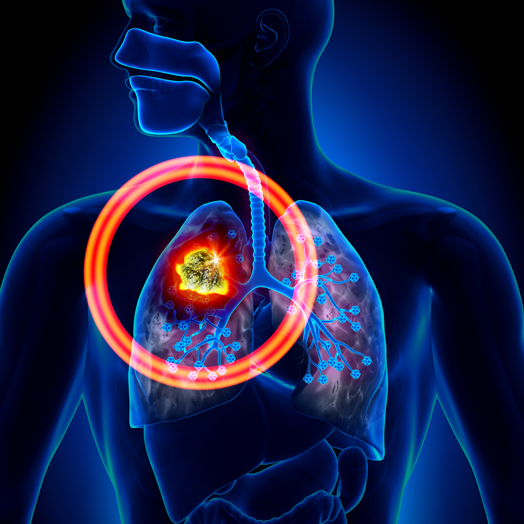
A biomarker is a biological characteristic that is objectively measured and evaluated as an indicator of normal or pathological processes. An imaging biomarker is a biological characteristic that is detectable on an image.
- Imaging biomarkers can be anatomical (i.e. the longest diameter of a large round lesion = structural information seen in the image”) or functional (i.e. looking for physiological aspects of the tumor in the image such as oxygenation levels, cellularity or vascularity)
- Imaging biomarkers can be qualitative (i.e., descriptive-“a nodule is present in the lung”) or quantitative (i.e., an objectively measured parameter-“the longest diameter of the nodule decreased by 5mm after treatment as compared with its size before treatment.”)
Several imaging biomarkers can contribute to one or several different response criteria.
Imaging biomarkers used by the RECIST (Response Evaluation Criteria in Solid Tumor) criteria:
- The first imaging biomarker used by RECIST is a quantitative one, which is the size of the longest axial diameter of target lesions.
- Another imaging biomarker used by RECIST is qualitative and records any appearance of new lesions: its evaluation is a binary and subjective outcome (“Is there a new lesion displayed somewhere in the image?”).
- The last imaging biomarker used in RECIST is also qualitative, and is the subjective evaluation of unequivocal progression of non-target lesions. (“Is there an obvious general increase of all these non-target lesions?”)
The therapeutic response per RECIST consists of the simultaneous monitoring of these three imaging biomarkers. In a clinical trial, the therapeutic tumor response can then be assessed on various imaging timepoints of a same patient using the RECIST criteria.
Read our blog: Medical Imaging Response Criteria, morphological measures
What are endpoints?
Endpoints, however, are the outcomes of a trial used to determine if a therapy is safe and effective. Once a patient reaches an endpoint, he/she is generally removed from the trial. There are different types of endpoints:
- Clinical endpoints include onset of toxicity, relapse or a hard endpoint such as patient death
- Surrogate endpoints are substitutes for a clinical endpoint and correlate with clinical benefit, such as Progression Free Survival. The most widely used clinical trial surrogate endpoint for Overall Survival (OS) is Progression Free Survival (PFS). PFS is defined as the time before patient experiences a progression per the response criteria or death. PFS is calculated as the median PFS time of the cohort after the end of the trial.
To which extent the reliability of imaging biomarkers evaluation has an impact on the reliability of the overall evaluation of clinical trial endpoints is a relevant question. That is the reason why imaging biomarkers, as root contributors to surrogate endpoints’ evaluations, are supposed to go through a rigorous qualification and validation process.
Historically, currently used imaging biomarkers did not undergo a stringent qualification process. This is the reason why in the last decade, several organizations started to define a specific qualification process for imaging biomarkers.
Aspects of the reliability of biomarkers that must be tested are:
- Accuracy: Performance in measuring what is really displayed in the image
- Precision: obtaining similar repeated measurements, without considering accuracy
- Normal patient variability: when measuring changes, to be able to discriminate between pathological changes from “normal” changes (from one human to another)
- Correlation to the disease: do we really measure a pathology? Is the lesion we see in the image really due to the disease or not?
- Usability: even if a biomarker encompasses all the above properties, its complexity of evaluation might preclude its adoption or its use in clinics.
In addition, some imaging biomarkers require the use of advanced software, like Median’s iSee® platform, such as imaging biomarkers that cannot be “seen” easily by the human eye. The best example would be texture biomarkers. Texture biomarkers are complex in nature and are extracted from the image using specific mathematical techniques and advanced software. Evaluating the texture biomarker would therefore require to also evaluate the techniques and advanced software used in the process.
Imaging biomarkers are the corner stone of modern radiology and imaging endpoints are a critical necessity to patient safety and understanding a drug’s effectiveness. They are major players in therapeutic decisions and drugs evaluations which is why they require advanced multidisciplinary expertise to provide the most accurate information possible.